Radiology of the Postoperative GI Tract PDF
Preview Radiology of the Postoperative GI Tract
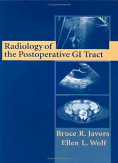
Radiology of the Postoperative GI Tract Springer New York Berlin Heidelberg Hong Kong London Milan Paris Tokyo Bruce R. Javors, MD Department of Radiology, Saint Vincent’s Hospital, New York, New York, USA Ellen L. Wolf, MD Department of Radiology, Montefiore Medical Center, Bronx, New York, USA Radiology of the Postoperative GI Tract With 320 Illustrations 1 3 Bruce R. Javors, MD Ellen L. Wolf, MD Department of Radiology Department of Radiology Saint Vincent’s Hospital Montefiore Medical Center New York, NY10003 Bronx, NY10467 USA USA [email protected] ewolf@montefiore.org Cover illustrations:Top:Intraoperative film from a cystic duct cholangiogram performed during a laparascopic cholecystectomy. Middle:Transverse sonogram demonstrates the encrusted tip of a ventriculoperitoneal shunt catheter surrounded by a sonolucent area with a hyperechoic rim. This represents a CSFpseudotumor or so-called “CSF’oma.” Bottom:CTsection through the mid-abdomen reveals a markedly dilated transverse duodenum in a patient who has undergone a Billroth II procedure (not shown). This is part of the spectrum of findings in afferent loop syndrome. Back cover:Frontal film from a double contrast upper GIseries shows a marginal ulcer in a patient who has under- gone a partial gastrectomy and gastrojejunostomy (Billroth IIprocedure). Library of Congress Cataloging-in-Publication Data Javors, Bruce R. Radiology of the postoperative GI tract / Bruce R. Javors, Ellen L. Wolf. p. ; cm. Includes bibliographical references and index. ISBN 0-387-95200-4 (h/c : alk.paper) 1. Gastrointestinal system—Radiography. 2. Gastrointestinal system—Surgery—Complications—Diagnosis. I. Wolf, Ellen L. II. Title. [DNLM: 1. Digestive System—radiography. 2. Digestive System Diseases—radiography. 3. Digestive System Surgical Procedures—adverse effects. 4. Postoperative Complications—radiography. WI 141 J4lr 2002] RC804.R6 J38 2002 617.4¢301—dc21 2002019467 ISBN 0-387-95200-4 Printed on acid-free paper. © 2003 Springer-Verlag New York, Inc. All rights reserved. This work may not be translated or copied in whole or in part without the written permission of the publisher (Springer-Verlag New York, Inc., 175 Fifth Avenue, New York, NY10010, USA), except for brief excerpts in connection with reviews or scholarly analysis. Use in connection with any form of information storage and retrieval, electronic adaptation, computer software, or by similar or dissimilar method- ology now known or hereafter developed is forbidden. The use in this publication of trade names, trademarks, service marks, and similar terms, even if they are not identified as such, is not to be taken as an expression of opinion as to whether or not they are subject to proprietary rights. While the advice and information in this book are believed to be true and accurate at the date of going to press, neither the authors nor the editors nor the publisher can accept any legal responsibility for any errors or omissions that may be made. The publisher makes no warranty, express or implied, with respect to the material contained herein. Printed in the United States of America. 9 8 7 6 5 4 3 2 1 SPIN 10790657 www.springer-ny.com Springer-Verlag New York Berlin Heidelberg Amember of BertelsmannSpringer Science+Business Media GmbH To my wife Susan and my daughter Alli, who have provided so much support and have enabled me to persevere during my travails. BRJ To my husband Gary, my daughters Bethany and Lesley, and to the memory of my parents, Isabel and Dr. Bernard S. Wolf ELW Preface The focus of this book is on the normal postoperative appearance of the gastrointestinal tract and the abnormalities specific to various surgical techniques. The authors have generally avoided the topics of abscesses, leaks, and other fluid collections unless specific attention was warranted. The imaging of recurrent neoplasm was also not included unless rather unique imaging characteristics were involved. The problem of recurrent Crohn’s disease was, however, included because of the controversies regarding its clinical and radiographic fea- tures. Organ transplantation was not addressed because such proce- dures are performed and followed at only a few centers. Over the last two decades the number of routine upper and lower gastrointestinal examinations has markedly decreased. More and more, the focus of conventional barium studies has shifted to the examina- tion of the postoperative patient. The last text dedicated to the post- operative appearance of the gastrointestinal tract was published more than 30 years ago, prior to the advent of computerized tomographic and magnetic resonance imaging. Although a few review articles and several chapters in major radiology textbooks have dealt with this subject, we felt there was a need for a more comprehensive approach. Our experience in teaching many residents has led us to realize that a knowledge of the surgical procedures themselves was at the heart of radiological comprehension, hence the emphasis on the basic prin- ciples of surgical technique in the chapters that follow. The line draw- ings that accompany the text were simplified to emphasize the anatomy and are not meant to be precise renditions of the actual surgical technique. Bruce R. Javors, MD Ellen L. Wolf, MD vii Acknowledgments The authors gratefully acknowledge the financial support of the E-Z- Em Corporation. Morton Meyers, MD, was instrumental in the birth of this book. Most of all we would like to thank our residents, who have over the years performed and monitored so many of the studies that follow. They have also asked the questions that pushed us to seek answers that at times were not readily available. From those basic ques- tions, the need for this book arose. We hope that what follows does justice to their quest for knowledge. We would like to thank Rob Albano at Springer-Verlag, for his patience. The authors would also like to thank Lourdes Guzman, Eleanor Murphy, and Seymour Sprayregen, MD, for their assistance. Bruce R. Javors, MD Ellen L. Wolf, MD ix Contents Preface . . . . . . . . . . . . . . . . . . . . . . . . . . . . . . . . . . . . . . . . . . . . . . vii Acknowledgments . . . . . . . . . . . . . . . . . . . . . . . . . . . . . . . . . . . . . ix Chapter 1 The General Abdomen . . . . . . . . . . . . . . . . . . . 1 Chapter 2 The Esophagus . . . . . . . . . . . . . . . . . . . . . . . . . . 61 Chapter 3 The Stomach . . . . . . . . . . . . . . . . . . . . . . . . . . . . . 137 Chapter 4 The Biliary Tree . . . . . . . . . . . . . . . . . . . . . . . . . . 225 Chapter 5 The Small Bowel . . . . . . . . . . . . . . . . . . . . . . . . . 279 Chapter 6 The Colon . . . . . . . . . . . . . . . . . . . . . . . . . . . . . . . 335 Index . . . . . . . . . . . . . . . . . . . . . . . . . . . . . . . . . . . . . . . . . . . . . . . 379 xi 1 The General Abdomen Free Intraperitoneal Air Free intraperitoneal air is expected in the immediate postoperative period. The frequency and duration of its detection may vary with the diagnostic modality utilized. The clinical importance of its detection may depend on whether the patient is receiving mechanical ventila- tion. Free intraperitoneal air may result from the dissection of air from a ruptured alveolus, back along the tracheobronchial tree, through the mediastinum, and either transdiaphragmaticly or via the retro- and subperitoneum into the abdominal spaces [1]. This can lead to a false positive diagnosis with resultant unnecessary exploration of the patient [1]. Free air may also result from peritoneal dialysis. Faulty bag exchange or exterior line exchange may allow air entry into the abdomen. Chest x-rays may show free air in 4% of patients [2]. However, up to 11% of cases of free air in patients undergoing peritoneal dialysis may be sec- ondary to gastrointestinal (GI) tract perforation. The amount of the air cannot be used to differentiate mechanical causes from pathological ones [2]. Conventional (plain film) radiographs of the abdomen may reveal free air in 30 to 77% of patients immediately after surgery [3–7]. This decreases to 38% on day 3 and 17% on day 17 [8]. The left lateral decu- bitus film, which allows air to rise and be highlighted between the liver and the parietal peritoneum, is positive in 53% of patients on post- operative day 3 and in 8% on day 6. As would be expected, these results compare unfavorably with the use of computerized tomography (CT) to detect free air. On postoperative day 3, 87% of CT images are posi- tive for free air, and fully half are positive on day 6 [8]. Serial exami- nations in 10 patients showed a decrease in the amount of free air in six patients during the same examination period, and a complete dis- appearance in the other four [8]. An area of some controversy is whether body habitus affects the rate at which free air is absorbed. Two older studies, based on plain films, detected a higher rate of free air in asthenic individuals [3,5]. Amore 1
Description: