Leaf anatomy of Cinnamomum schaeffer (Lauraceae) with special reference to oil and mucilage cells PDF
Preview Leaf anatomy of Cinnamomum schaeffer (Lauraceae) with special reference to oil and mucilage cells
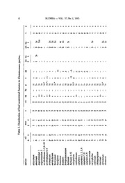
BLUMEA 37 (1992) 1-30 Leaf anatomy of Cinnamomum schaeffer (Lauraceae) with special reference to oil and mucilage cells M.E. Bakker A.F.Gerritsen & P.J. van der Schaaf Rijksherbarium/HortusBotanicus,Leiden,The Netherlands Summary Themorphologyanddistribution patternsofoiland mucilagecells inthe leafof 150species ofCin- namomum aredescribed. Idioblastsarealways presentin thepalisadeandthe spongy parenchyma. Usually both oiland mucilagecells occur; in somespecies eitheroil ormucilagecells arepresent. Bothtypes ofidioblastspossess asuberized wall layer. The idioblastsvary between species in size/ shape,stainabilityand number. Variations in the distributionpatterncanpartly beexplainedby the proposedhomologyofthe oil andmucilagecells. Otherleafanatomicalcharactersarealso mentioned,such aslamina andcuticle thickness,bundle sheath extensions,sclerification ofthe epidermal,thepalisade, and the spongy parenchymacells, number ofpalisadelayers, presenceorabsence ofa hypodermis,the indumentum and papillate ab- axialepidermalcells,andthe venationpattern. Mostspecies differfrom each other in onlyoneorfew leafanatomical characters (includingoil andmucilagecells). Agreatmany combinations incharacter distribution were observed. However, the distribution patterninapproximately one-fifth ofall studied species deviated largely from the typical leaf anatomical character distribution patternoccurring in the majority ofCinnamomum species.There wasamaximumofseven differingfeaturesoutofthesixteen features studied. Within thisgroup almost allneotropicalCinnamomum species are included. The latterspecies lack scleri- fiedepidermalcells and almostall have penninervedinstead ofthe generallyoccurring triplinerved leaves. Clusteranalysesbased on all leafanatomical features studied revealed that thedistribution pat- terns oftheoiland mucilagecells play asignificant partin the groupingofthe species. Therefore oil and mucilagecells possessatleast somediagnosticvaluewithin thegenus Cinnamomum.The systematic significanceofoil andmucilagecells atthe infragenericlevel remains uncertain forlack ofadetailed infragenericclassification ofCinnamomum for comparisonwith the idioblast distribu- tion patterns. Introduction Inaprevious study onoiland mucilage cellsin species ofthegenusAnnona(Bakker & Gerritsen, 1992) we explored the systematic valueoftheseidioblasts.Since the distributionoftheidioblastsshowedlarge variationwithingroupsofspecies it was concludedthat oil and mucilage cells had littleor no taxonomic valuebelow the genuslevel.Thiscan partly be understoodin thelightoftheproposed homology of oiland mucilage cells(Bakker & Gerritsen, 1989, 1990, 1992;Bakker etal., 1991). 2 BLUMEA VOL. 37, No. 1, 1992 Inthepresentpaperthe diagnostic (discriminating) andsystematic valueofoiland mucilagecellswillbeinvestigated forthegenusCinnamomumSchaeffer. Cinnamomumis a genus withabout 200accepted species (Kostermans, 1964, 1986,1988). Anumberofneotropical species previously placed inothergenerasuch as Oreodaphne,Persea,Laurus, Ocotea,andPhoebehavebeen transferredtoCinna- momum (Kostermans, 1961, 1988;Van derWerff, 1991a,b). The not neotropical species arepresent in the Malesian area, Australia, Asia, and the Pacific Islands (Kostermans, 1961, 1968, 1969, 1970a, b, 1983, 1986, 1988; Hyland, 1989).The genusis especially known for theproduction ofcinnamonandcamphor (Pax, 1889; Hegnauer, 1966; Kostermans, 1986).Theuseofthebark(cinnamon) datesfroman- cienttimes(seeKostermans, 1986). Nowadays, theplantsarealso usedas medicine andinthecosmeticandperfume industry. Leafanatomicaldescriptions ofCinnamomumspecies are scarce. General notes were given by Pax (1889), Solereder(1899), Metcalfe& Chalk(1950) andMetcalfe (1987). Perrot (1891) describedthehistology ofoil and mucilage cellsin species of Lauraceae,including five Cinnamomumspecies. Lateronly two detailedleafana- tomical studies were published for arestricted numberof Cinnamomumspecies (Santos, 1930; Marlier-Spirlet, 1946). ThegenusCinnamomumis poorly understoodand poses many taxonomicdiffi- culties. Thetwo sections distinguished by Meissner (1864): MalabathrumMeissn. and Camphora Nees, are still accepted nowadays as the only subdivision ofthe genus(Kostermans, 1986).The sections, besidestheir differentsmells, also canbe distinguished by budmorphology andleafarrangement(Kostermans, 1986). In thepresentpaperthedistributionofoil andmucilage cells andotherleafana- tomicalfeaturesare describedfor 150species ofCinnamomum.Acomparison ofthe leafanatomicalcharacterdistributioninthespecies is carried out by meansofcluster analysis andtheresults are discussed inthelightoftheexisting classificationin order to evaluatethe diagnostic and/orsystematic valueoftheoiland mucilage cellsbelow thegenuslevel. MATERIALAND METHODS Materialand fixation The207specimens ofthe 150speciesofCinnamomumstudiedare listedinTable 1. Inthis study leavesfromherbariumvouchers, kept in Leiden, wereboiledinwater untilthey permanently sank inwater, andthenwere storedin aFAPA-solution. Fresh leavesofC. burmanni, C. camphora, C. daphnoides, C. iners, C. kina- baluense, C. loureirii, C. obtusifolium, C.parthenoxylon, C. sericeum, C. tamala, C. verum, and C. spec., grown in greenhouses of several Botanical Gardens in Western Europe (in Table 1 marked with an asterisk), were collected and fixed inFAPA-solution. Fresh leaves from C. burmanni(Leiden, Java), C. camphora (Berkeley, Bogor), C. iners (Wageningen, Bogor), Ci . kinabaluense(Bogor), C. parthenoxylon andC. verum(Delft) were fixedinmodifiedKarnovsky fixative fol- lowedby OSO4 andembeddedinEpon (see Bakker& Gerritsen, 1989). M.E.Bakker, A.F.Gerritsen & P.J.van derSchaaf: Leafanatomy of Cinnamomum 3 Table 1. List ofmaterialexaminedofCinnamomum. All materialis herbariummaterial locatedin L. altissimum Kosterm.: Chelliah FRI 6543, Malaysia —amoenum(Nees) Kosterm.: Smith & Klein 11260, Brazil —angustitepalum Kosterm. 1: Kostermans 12974,Borneo —angusti- tepalum 2: Brain anak Tada S 16253,Borneo — archboldianum Allen 1: Craven & Schodde 1239,New Guinea —archboldianum 2: bb. 25066,New Guinea — archboldianum 3: Koster BW6854,New Guinea —archboldianum 4: Streimann & Stevens LAE 54854, New Guinea — archboldianum 5: Foreman & Vinas LAE 60127, New Guinea — archboldianum 6: Schodde & Craven 5014,New Guinea — archboldianum 7: Aet& Idjan 394,New Guinea —archbold- ianum 8: Carr 14814,PapuaNew Guinea —archboldianum9:Carr 13253,PapuaNew Guinea —archboldianum 10:StevensLAE54769,NewGuinea—- aromaticumNees: Schewes.n.,s.l.— aureo-fulvumGamble: Wong Khoon MengFRI 32230, Malaysia —baileyanum (F. Muell.) Francis: Hyland 7547, Australia —bejolghota (Ham.) Sweet: Schewe s.n., Asia —bintulense Kosterm.:DingHou349,Borneo—bodinieri Gamble/Lev.:Beauvais 132,China —brevipedun- culatumChang:Ching-enChang6643,Taiwan—bullatum Kosterm.:WomersleyNGF11335,New Guinea —burmanni (Nees) Blume 1: Schewe s.n., Japan—burmanni 2: de Vriese & Teijs- mann s.n., Sumatra—burmanni 3: de Vriese & Teijsmann s.n., Java—burmanni 4: Boer- lage 1888,Java —burmanni 5: Keith For.Dep. No. 5999, Borneo —burmanni 6: Perrottet s.n., Philippines — burmanni 7: Teijsmann s.n., Celebes — burmanni 8: Schmutz 2836, Lesser Sunda Islands —burmanni 9: Iboet514, Lesser Sunda Islands —burmanni 10: Kos- termans s.n.,Java—burmanni 11*: Hortus Botanicus Leiden,The Netherlands —burmanni 12*: Baas s.n.,Cibodas, Java — calciphilumKosterm.: Paul et al. S 37382,Borneo —cam- bodianum Lec.: Poilane 23247, Cambodia —camphora(L.) Presl 1: d'Aleizette 558m,Japan — camphora2*: Bot. Gardens Kew No. 00073 12261,U.K. — camphora 3*: Royal Bot. Garden EdinburghNo. 69 6417, U.K.—camphora4*: Bot. Garden Techn. Univ. Delft, The Netherlands — camphora5*: Bot. Garden Utrecht No. 73GR00538,The Netherlands —cam- phora6*: Van der Werffs.n.,Berkeley, U.S.A.—camphora7*: Bot.Garden BogorNo. XXB 112,Java — camphora 2*: Baas s.n.,Cibodas,Java —camphora variegata 9*: Bot. Gardens KewNo. 00073 12262,U.K.—cappara-corondeBlume: Kostermans25596,Ceylon—caryo- phyllus (Lour.) Moore: Bordeneuve (Chevalier40969),Indochina —celebicum Miq.: bb. 5555, Celebes —chittagongenseKosterm.: King249,India —cinereum Gamble: Corner s.n., Ma- laysia —citriodorum Thw.: Kostermans 23455,Ceylon—clemensii Allen: Eyma5093,New Guinea —cordatum Kosterm.: Whitmore FRI20470, Malaysia —coriaceum Cammerl.: bb. 14079,Borneo —corneriKosterm.: Carson SAN 28012,Borneo —crassinervium Miq.:Puasa, B.N.B.For. Dep. 3159,Borneo —crispulumKosterm.: Poilane21913, Indochina —cubense (Nees) Kosterm.: Sintenis 1036,Puerto Rico —culitlawan (L.) Kosterm.: Labill.,Blackw.t.? 391,Moluccas —curvifolium (Lour.) Nees: Poilane 32.126,Indochina —cuspidatumMiq. 1: SuppiahFRI 11901,Malaysia—cuspidatum 2: de Wilde & de Wilde-Duyfjes 12889,Sumatra — damhaenseKosterm.:Chevalier 37468,Asia—daphnoidesSieb. & Zucc. 1:Koidzumi s.n., Eur./Asia— daphnoides2*: Bot. Gardens Kew No. 00073 12263,U.K. —deschampsii Gamble: Corner SFN 31000,Singapore — dewildei Kosterm.: bb. 2796, Sumatra— doeder- leinii Engl.: Fosberg 37185,Ryukyu Islands — dubium Nees 1: Kostermans 24095, Ceylon — dubium 2: s.n., Java— durifoliumKosterm.: Poilane 9030, Asia — ebaloi Kosterm.: Ebalo 1198,Philippines—effusum (Meissn.) Kosterm.: Sousa 7876, Mexico—elephantinumKos- term.: B6jaud 742m,Cambodia— ellipticifolium Kosterm.: Balus? 12288, Burma — elon- gatum (Vahl)Kosterm.:Curtiss 309, WestIndies (Central America) —englerianum Schewe: Ledermann 9806, New Guinea —eugenoliferumKosterm.: bb. 32907,New Guinea —fouil- 4 BLUMEA VOL. 37,No. 1, 1992 (Table 1continued) loyiKosterm.: Endert3840,Borneo —frodiniiKosterm.: Henty &KatikNGF49456,NewGuinea —gigaphyllumKosterm.: TelussaBW5161,New Guinea—glaucescens(Wall.) Drury:Poilane 1272,Asia—goaense Kosterm.: Stocks s.n.,Asia—grandiflorumKosterm.: Hoogland5052, New Guinea — griffithii Meissn. 1: J.C. 1641,Malaysia — griffithii 2: Kostermans 5283, Borneo —iners Reinw./Blume 1: Pierre 5170,Asia —iners 2: Kostermans? s.n., Java — iners 3*: Bot. Gard. Agricult. Univ. WageningenNo. 72 PTO 1275,The Netherlands —iners 4*: Bot. Gard. Bogor No. XXB74, Java—insulari-montanum Hay.: Kao 9834,Taiwan — javanicum Blume 1: bb. 2729, Sumatra —javanicum2: s.n.,Java —kami Kosterm.: Frodin NGF28255,New Guinea —keralaense Kosterm.:Kostermans 26283,South India—kinabalu- ense Heine 1:Aban GibotSAN66838,Borneo—kinabaluense2*: Bot.Gard. BogorNo. XXA 85,Java—kunstleriRidley: Poilane 31019,Indochina—laubatii F.Muell.: Irvine 1668,Aus- tralia —ledermanniiSchewe: Powell UPNG 1639,PapuaNew Guinea —litseafoliumThw.: Davidse 8455, Ceylon — longitubumKosterm.: Poilane 29260, Asia — loureiriiNees 1: Yoshida 2201,Japan—loureirii2*:Royal Bot.Gard.EdinburghNo. 100029,U.K.—lucens Miq.: Meebold 14876,Burma —macrocarpum Hook, f.:Kostermans 24534, South India — macrophyllum Miq.: Kostermans 1230,Moluccas —magnifolium Kosterm.: Poilane 29510, Indochina —maireiLev.: s.n.,Asia—malabaricum Garc.?: Ridsdale 495,Asia—malabath- rum (Burm.f.) Blume: Kostermans 26119, South India —malayanum Kosterm.: Kochummen FRI 16657,Malaysia —melastomaceum Kosterm.: Poilane 29636, Indochina — melliodo- rum Kosterm.: Sayers NGF24238,New Guinea —mendozae Kosterm.: MendozaPNH 42310, Philippines —mercadoi Vid.: Merrill Sp. Blanc. No. 758,Philippines —microcarpum Kos- term.: Martin S 38157,Borneo —microphyllum Ridley: Whitmore FRI 15582, Malaysia— mollissimum Hook, f.:Corner SFN 30885, Malaysia —montanum (Sw.) Berchth. & Presl: Krug& Urban 5259,Jamaica—myrianthum Merr.:Conklin PNH 37897,Philippines —myr- tifoliumKosterm.: Chevalier 35.993, Cambodia — nalingway Kosterm.: Kostermans 270, Asia—nooteboomii Kosterm.: bb. 6628, Sumatra —obtusifoliumNees *: Royal Bot. Gard. EdinburghNo. 350159,U.K.—oliveri F.M.Bailey: Tadman s.n.,Australia—-osmeophleum Kan.: Kao 9650, Taiwan —ovalifoliumWight: Hoogland 11550,Ceylon— ovatum Allen: Jean 38.397,Indochina —pachyphyllum Kosterm.: Sinclair & Kish bin Salleh SFN 39922, Malaysia —paieiKosterm.: Paie & Mamit S29332, Borneo—panayense Kosterm.: Martelino & Edafio BS 35653,Philippines—parthenoxylon Meissner *: Bot. Gard.BogorNo. XXB41, Java —perrottetiiMeissn.: Kostermans 25803, India —piniodorum Schewe: Versteegh BW 3919, New Guinea —podagricum Kosterm.: Sayers NGF 21699, New Guinea—poilanei Kosterm.: Poilane 33772, Indochina —politumMiq.: Smythies S 7803, Borneo —polyadel- phum (Lour.)Kosterm.: Poilane 28504,Indochina—porphyrospermumKosterm.: de Wilde & de Wilde-Duyfjes 12984,Sumatra —porrectum (Roxb.)Kosterm. 1:Bodinier 1118,China — porrectum2: Burkill KEP 76692,Malaysia—porrectum3: Rahmat siToroes 4694, Sumatra —propinquumF.M. Bailey: Hyland 6577, Australia —pseudopedunculatumHay.: Tuyama s.n.,Bonin Island —psychotrioides (H.B.K.) Kosterm.: Tokatiaa/1541,Mexico—racemo- sumKosterm.: Shea & Minjuk SAN76182, Borneo —reticulatum Hay.: Chang 3771,Taiwan —rhynchophyllum Miq. 1: Thorenaar 52, Sumatra —rhynchophyllum2: Clemens 22194, Borneo —rhynchophyllum 3: Sinanggui SAN 56217, Borneo —rhynchophyllum 4: Gibot SAN 37019, Borneo — rhynchophyllum5: Kostermans 4559, Borneo —rhynchophyllum 6: de Wilde & de Wilde-Duyfjes 15545,Sumatra—rhynchophyllum7: Forbes 2969, Sumatra —rhynchophyllum8: Ogata KEP 105031,Malaysia — rhynchophyllum9: Grashoff 733, Sumatra—riedelianum Kosterm.: Riedel s.n., Brazil —rigidum Gillespie: Webster & Hild- roth 14198,Fiji — riparium Gamble:Ridsdale 628, South India—rivulorumKosterm.: Kos- termans 27831,Ceylon —rupestre Kosterm.: Ridsdale SMHI 264,Philippines— scalarinerve M.E.Bakker, A.F.Gerritsen & P.J.van derSchaaf: Leafanatomy of Cinnamomum 5 (Table 1continued) Kosterm.:Chevalier 38.371,Indochina —scortechinii Gamble: Poilane 11143,Asia—sellowi- anum(Nees& Mart.)Kosterm.:Hatschbach 17399,Brazil—sericans Hance:Pierre5170,Asia— sericeum Siebold *: Royal Bot. Gard. EdinburghNo. 69 6418, U.K. —sessilifolium Kan.: Hosakawa 5954,Ponape.Pacific — simondii Lec.:Chevalier 32075, Indochina — sinharajense Kosterm.:Kostermans27486,Ceylon—sintok Blume: Kostermans s.n., Java—solomonense Allen: Waterhouse 129A-B,Solomon Islands —soncaurium (Ham.)Kosterm.: Hansen & Smiti- nand 12882,Thailand —spec.*:Royal Bot.Gard. EdinburghNo. 69 6352,U.K.—subavenop- sisKosterm.:BoschproefstationCel/IV-94,Celebes —subavenium Miq.1:Put3526,Thailand —subavenium 2:Kostermans37,Sumatra—subcuneatum Miq.:Lorzing 11289,Sumatra— subpenninervumKosterm.: Poilane9680, Asia —sulphuratumNees: Hook. f. & Thomson 2617,Asia—tahyanumKosterm.:Othman etal.S 41520,Borneo—tamala (Ham.)Th.Nees& Eberm. 1: Tatemi Shimizu etal. T-20932,Thailand —tamala 2*: Bot. Gardens Kew No. 00073 12264,U.K.—tampicense(Meissn.) Kosterm.:s.n.,Mexico—tazia'Hamilton' Hook,f.:Bot. Garden Calcutta s.n.,Asia —tepalinumKosterm.: Koelz29958, India —tonkinense (Lec.) Chev.: Poilane 32261, Indochina — trichophyllumQuis. & Merr.: Meijer 9759,Celebes — triplinerve (R. & P.) Kosterm.: Dombey 201, Peru —tsoi Allen: Poilane 24801, Annam — vaccinifolium Kosterm.: Sleumer & Vink BW 14254,New Guinea — velutinum Ridley: WhitmoreFRI12116,Malaysia—verumPresl1:Hasskarl s.n.,Japan—verum 2:Wight2511, India— verum 3:Sedies? s.n.,Mauretania — verum 4*: Bot. Gard. Berlin-Dahlem,Germany— verum 5*: Bot. Gard. Techn. Univ. Delft,The Netherlands — verum6*: Bot.Gard. Agric. Univ. Wageningen,The Netherlands — vesiculosum (Nees)Kosterm.: Klein 3.269, Brazil — vimi- neum Nees: LohFRI 19170,Malaysia —virens R.T. Baker: Jones 2693, Australia — wightii Meissn.: Kostermans 25731,India —xanthoneurum Blume: Versteegh& Vink BW 8309,New Guinea — xylophyllum Kosterm.: Kostermans 12981,Borneo —zollingerii (de Lukm.) Kos- term. 1: Walker et al. 6059,Okinawa Island —zollingerii 2:Fosberg 38155, Ryukyu Islands. Sectioning, staining and microscopy Themethods usedforsectioning andstaining oftheleavesare identicalto those describedforAnnonaleaves inBakker& Gerritsen (1992). Thick(30 pm) transverse sections ofFAPA-fixed leaves were stained foroil with Sudan IV or Chrysoidin/ Acridinred. Mucilage was stainedwith AlcianBlue. For suberinthe Berberinand AnilinBlue fluorescentstaining methodwas applied. Ultrathinand 1 pm sections ofEpon-embedded materialwereexaminedwithtransmissionelectron microscopy (TEM) andlightmicroscopy (LM)respectively (see Bakker&Gerritsen, 1992;Bakker et al., 1991). Someleaffragments were air-dried, gold-sputtered andexaminedwiththescan- ningelectronmicroscope (SEM). Examination and measurements Inthe30pmsectionsthenumberofoiland/ormucilage cellsinthepalisade and/ or spongyparenchyma was countedand calculatedpermmleafwidth. In thesame sectionsthelaminathickness was measuredandthenumber(per mmleafwidth)and length ofhairs was determined(see Bakker& Gerritsen, 1992). Thepresence/ab- senceofanindumentumwas verifiedby examinationoftheleaveswithastereo mi- croscopeandpartly withascanning electronmicroscope (SEM). 6 BLUMEA VOL. 37, No. 1, 1992 Phenetic analysis Phenetic analyses were carried out with the help of NTSYS-pc, version 1.30 (Rohlf, 1986),acomputerprogramfornumericaltaxonomicandmultivariateanaly- ses. Eachanalysis consistsofa two-stepprocedure,i.e.,thecomputation oftheover- allsimilarityamong taxa and therepresentation thereofby aphenogram. Inthefirst step a datamatrix, basedon Table2, is usedto calculatethe (dis)similarity among taxa expressed as Manhattandistances.Inthesecond step,creating aphenogram, the pairwise distances amongtaxa serve as input fortheanalysis, using UPGMA (Un- weighed Pair-Group MethodusingarithmeticAverages) asa clustercriterion. RESULTS Oil and mucilage cells 30 pm sections - Light microscopy SudanIV— Oil cellswererecognized bythestainedcontents (red, orange, pink, brown; depending onthe species). Theoil is presentas small irregularbodies (Fig. 1A),clustering globules (Fig. IB), a distinctly stainedmass (Fig. 1C),or ahomo- geneousoil drop.Theoil dropmay be visibly attachedto thewallby acupule (Fig. ID). Otheroil cellsdid notcontainoil but couldbe distinguished bythepresenceofa cupule (Fig. IE) and/ora characteristicfoldin thecell wall (Fig. IE, F). Theseoil cellshad a glossy pink appearance. Mucilage cells were unstainedandempty. The suberizedlayer was visibleas athinred lineintheoil andmucilage cells. Chrysoidinlacridin red — Theoil cells wererecognized as describedforthe SudanIV-staining, butgenerally theidioblastswere less clearbecauseofthestrong background staining. Mucilage cells wereunstained(empty) or faintly yellowish. AlcianBlue— Oil cellsstainedglossy greenish-blue or wereempty(Fig.2A) and couldbedistinguished fromthemucilage cellsby athickdarkbluecellwall, sometimes containing acupule, or by thering-like appearanceofthecytoplasm. Inmucilage cells theretainedmucilage distinctly stainedblueandsometimesextruded fromthecells (Fig. 2A, B,D). Themucilageoftenshowed(circular) striation(Fig. 2C). Thepresence ofelderpith fragments (usedto clamp leaffragmentsforsectioning) stickingto the sectionsalsoindicatedthepresenceofmucilage intheleaf.In controlsamples themuci- lage was stained less densely and generally spread out over thesectionas aresultof thedissolutionofthemucilage inwater, thereby obscuring themesophyll (Fig. 2D). 30 pm sections - Fluorescence microscopy Thecell wallsofoiland mucilage cellsshowedautofluorescenceofthesuberized walllayer. Whenstainedspecifically forsuberin (Berberin) thislayer appeared as a thinyellow/whitish thinline inthecellwall. Thisstaining reactionhasbeen describ- edearlierforoiland mucilagecellsofCinnamomumburmanni(Bakker et al„ 1991). 1pm sections - Light microscopy Both idioblast types were easily distinguished fromeach other by theiroverall cytoplasmic composition. Oil cells were also characterized by the presence ofa cupule. Mucilage cells showedthe distinctly purple-stained mucilage and/or the characteristicpattern ofthe cytoplasmic strands. M.E.Bakker, A.F.Gerritsen & P.J.vander Schaaf: Leafanatomy of Cinnamomum 7 Transmission electron microscopy The suberizedwall layer was already observedagainst theinner sideofthe cell wallin a youngoilcell inthe leafofCinnamomumcamphora (Fig. 3A) andturned outtobealways presentin mature oil(Fig. 3D)and mucilagecells. Thecomposition ofthecytoplasm oftheoiland mucilage cells intheleafis very similarto thatde- scribedfortheidioblastsintheshootapexofC.burmanni(Bakker& Gerritsen, 1989; Bakker et al., 1991). In a very youngoil cell, present in thepalisade parenchyma, electron denseoil dropletsand characteristicplastids werepresentinthecytoplasm (Fig. 3B). Theplastids lacked thylakoids but containedstarch, dark globules and very smallwhiteglobules (Fig. 3B). Inanotheryoungoil cellinthepalisade paren- chyma athickenedpartoftheinnerwalllayerwas observed: thecupule base(Fig. 3C). Inmature oilcellsinleavesofdifferentCinnamomumspecies acupule was generally observed(Fig. 3D), showing the cupule basefrom which the cupule projects into the cytoplasm surrounding theoilcavity. These observations are identicalto those describedearlierforcupules in oilcellsofC. burmanni(Bakker& Gerritsen, 1989; Bakker etal., 1991) andAnnona muricata(Bakker & Gerritsen, 1990). Shape and size In mostspecies the oil cells are oblong-ovoid inthepalisade parenchyma (Fig. 1A,C,D) andmoreor less globularin thespongyparenchyma (Fig. IB, E, F). The mucilage cells generally areovoidin thepalisade parenchyma (Fig. 2A,B, D) and globularin thespongyparenchyma (Fig. 2B, C). Thesizes oftheidioblasts varyper species (Figs. 1A-F& 2A-D). In general mucilagecellsare largerthanoilcells. Distribution Oil and/ormucilage cellsare always presentinboththepalisade andthespongy parenchyma (Table 2). Mostspecies possessboth oilandmucilage cellsin theleaves. The majorityofthese species containthetwo idioblasttypesin boththepalisade and thespongyparenchyma. Theexclusivepresence ofoil cellsintheleafoccurred inc. 17% ofthespecies and only 5 species containedexclusively mucilage cellsin the leaves (Cinnamomumarchboldianum[no.4and 5], C. magnifolium, C. nalingway, C. rivulorumandC.sinharajense). Inthespongyparenchyma idioblastsare usually locatedagainstthelowersideof thepalisadeparenchyma (Fig. 2A, B)and againsttheabaxialepidermal cells. Of 18 species ofwhich morethan 1 specimen was studied 11 were constant for thedistributionoftheoil and mucilagecells inthepalisade andspongyparenchyma (C. angustitepalum [2 specimens], C. burmanni[12], C.cuspidatum [2], C. daph- noides [2], C. javanicum [2], C. loureirii[2], C.porrectum[3], C. rhynchophyllum [9], C.subavenium[2], C. tamala[2] and C. verum [6]). Theotherspecies showed variationintheirdistributionpatterns. Smallvariationsconcern thepresence/absence ofoilor mucilagecells inoneofthemesophyll layers (C. griffithii[2]andC. zollin- gerii [2]). Larger variationsoccur whenpartofthespecimens lacks one typeofidio- blast iC. archboldianum[10], C. camphora [9], C. dubium[2] and C. iners [4]). Cinnnamomumkinabaluense[2] showedtwodifferentdistributionpatterns(Table 2). Thefrequency oftheidioblast typesin theleafwas foundtobe highlyvariable betweenspecies (Table 2). Betweenspecimens ofthesame species only minorvari- ations occurred in thenumberofidioblasts.Themaximumnumberin the palisade 8 BLUMEA VOL. 37, No. 1, 1992 M.E. Bakker,A.F.Gerritsen &P.J. vander Schaaf: Leaf anatomy ofCinnamomum 9 Fig. 2.Transverse 30µm thicksections ofCinnamomum species. A—D:Mucilagecells stained with Alcian Blue. Lightmicroscopy. All x 345 —A:C. virens. Mucilagecell in thepalisadeparen- chymashowingextrusion ofthe stained mucilagefrom the cell into another cell. Also notethe neighbouringunstained oilcell (oc) in thespongy parenchyma.—B: C.nalingway. Mucilagecells in thepalisadeand spongy parenchyma,both extrudingmucilageintothe intercellular spacesofthe spongy parenchyma.Notethe darker stainedmucilagein the centre ofthe cells.—C:C. vaccini- folium. Mucilagecellin thespongy parenchyma.Note the circularstriations in the mucilage.—D: C.magnifolium. Mucilagecell in thepalisadeparenchyma.Note the less denselystained mucilage extrudedfromthe cellandspreadoutover thesection. Fig. 1. Transverse 30 µm thick sections of Cinnamomum species. A—F: Oil cells stained with Sudan IV. Light microscopy. All x 725.— A: C. cubense. Oil cell in the palisadeparenchyma showingloosepieces ofstainedoil. —B: C. crispulum. Oil cell in the spongy parenchymafilled withamass of globulesofstainedoil. —C: C. vesiculosum.Oil cell in the lowerlayerofthepali- sadeparenchymafilledwithaheterogeneousoildrop.—D: C. tampicense.Oil cellin thepalisade parenchymafilledwith alargeoildrop.—E:C.brevipedunculatum.Empty oilcellin the spongy parenchymashowingacupule(arrow)and afoldin thecell wall(arrowhead).—F:C. vimineum.Large emptyoilcell in the spongy parenchymashowingthe foldin the cell wall. 10 BLUMEA VOL. 37, No. 1, 1992 Fig.3.Cinnamomum camphora 8. A—D:Transmission electron microscopy.— A: Detail ofthe cellwall ofa very youngoilcellinwhich only asuberized wall layer(s) has been depositedbetweentheprimary cellwall(w) andthe cytoplasm(c); X58,200.—B:Detail ofthe cytoplasmofyoungoilcelldepictedin A.Note the specificplastids containingstarch (st), dark globules(arrowheads),andsmall whiteglobules (smallarrows).The plastidslack thylakoids. Notethelargeoil globule(o)inthe cytoplasm;X 27,250. — C:Detail ofthecell wall ofa young oil cellshowing the developmentofacupulebase (cb) deposited againstthesuberized wall layer(s). The cupulebaseis thethickened part oftheinner walllayerwhichis depositedagainstthe suberizedlayerthroughoutthewholeoilcell; X 27,250.— D:Detail ofadeveloped cupuleinanolderoilcell inthe palisadeparenchyma. Notethe cupulebase (cb) from whichthe cupule proper (arrows)projectsinto the cytoplasmand surrounds theoilcavity (oc); x 16,930. Fig.4.Transverse 30µm thicksections ofleaves ofCinnamomumspecies.A—F: Stained withSudan IV. Lightmicroscopy. —A:C.vaccinifolium.Extremelythick,denselystainedcuticle(17µm)showinganirreg- ularoutersurface;x400.—B:C.glaucescens. Adaxialepidermis withlargenon-sclerifiedepidermalcells. Notethe cutinized(stained) anticlinalwalls;x 400. —C:C. wightii. Vertically transcurrentlowerorder veinwithsclerenchymatousbundlesheathextensions; x 400.—D:C. velutinum. Detail oftheuppermost part ofa verticallytranscurrent lowerorder vein showingthe continuation ofthe sclerified bundle sheath cellswiththesclerified adaxialepidermalcells;x 1,000.— E:C. sintok. Layer ofalignedsclerified outer periclinal wallsofthepalisadecellsunderneaththe smallepidermalcells;x 400.— F:C. sericeum.Hypo- dermis(hy) underneaththeadaxialepidermis.Note thesclerenchymatouscellsinthislayer(arrow); x400.
