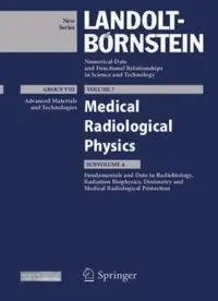
Fundamentals and Data in Radiobiology, Radiation Biophysics, Dosimetry and Medical Radiological Protection PDF
Preview Fundamentals and Data in Radiobiology, Radiation Biophysics, Dosimetry and Medical Radiological Protection
New Series Numerical Data and Functional Relationships in Science and Technology GROUP VIII VOLUME 7 Advanced Materials Medical and Technologies Radiological Physics SUBVOLUME A Fundamentals and Data in Radiobiology, Radiation Biophysics, Dosimetry and Medical Radiological Protection 123 Landolt-Börnstein Numerical Data and Functional Relationships in Science and Technology New Series Group VIII: Advanced Materials and Technologies Volume 7 Medical Radiological Physics Subvolume A Fundamentals and Data in Radiobiology, Radiation Biophysics, Dosimetry and Medical Radiological Protection A. Almén, J.H. Bernhardt, L. Johansson, K.-U. Kasch, A. Kaul, H.-M. Kramer, S. Mattsson, B.M. Moores, D. Noßke, H.-J. Selbach, F.-E. Stieve, J. Valentin, S. Vatnitsky Edited by A. Kaul ISSN 1619-4802 (Advanced Materials and Technologies) ISBN 978-3-642-23683-9 Springer Berlin Heidelberg New York Library of Congress Cataloging in Publication Data Zahlenwerte und Funktionen aus Naturwissenschaften und Technik, Neue Serie Vol. VIII/7A: Editor: A. Kaul At head of title: Landolt-Börnstein. Added t.p.: Numerical data and functional relationships in science and technology. Tables chiefly in English. Intended to supersede the Physikalisch-chemische Tabellen by H. Landolt and R. Börnstein of which the 6th ed. began publication in 1950 under title: Zahlenwerte und Funktionen aus Physik, Chemie, Astronomie, Geophysik und Technik. Vols. published after v. 1 of group I have imprint: Berlin, New York, Springer-Verlag Includes bibliographies. 1. Physics--Tables. 2. Chemistry--Tables. 3. Engineering--Tables. I. Börnstein, R. (Richard), 1852-1913. II. Landolt, H. (Hans), 1831-1910. III. Physikalisch-chemische Tabellen. IV. Title: Numerical data and functional relationships in science and technology. QC61.23 502'.12 62-53136 This work is subject to copyright. All rights are reserved, whether the whole or part of the material is concerned, specifically the rights of translation, reprinting, reuse of illustrations, recitation, broadcasting, reproduction on microfilm or in other ways, and storage in data banks. Duplication of this publication or parts thereof is permitted only under the provisions of the German Copyright Law of September 9, 1965, in its current version, and permission for use must always be obtained from Springer-Verlag. Violations are liable for prosecution act under German Copyright Law. Springer is a part of Springer Science+Business Media springeronline.com © Springer-Verlag Berlin Heidelberg 2012 Printed in Germany The use of general descriptive names, registered names, trademarks, etc. in this publication does not imply, even in the absence of a specific statement, that such names are exempt from the relevant protective laws and regulations and therefore free for general use. Product Liability: The data and other information in this handbook have been carefully extracted and evaluated by experts from the original literature. Furthermore, they have been checked for correctness by authors and the editorial staff before printing. Nevertheless, the publisher can give no guarantee for the correctness of the data and information provided. In any individual case of application, the respective user must check the correctness by consulting other relevant sources of information. Cover layout: Erich Kirchner, Heidelberg Typesetting: Authors and Redaktion Landolt-Börnstein, Heidelberg SPIN: 1174 1213 63/3020 - 5 4 3 2 1 0 – Printed on acid-free paper Editor A. Kaul Okerstraße 13 38300 Wolfenbüttel, Germany e-mail: [email protected] formerly Bundesamt für Strahlenschutz Willy-Brandt-Straße 5 38226 Salzgitter, Germany and Physikalisch-Technische Bundesanstalt Bundesallee 100 38116 Braunschweig, Germany Authors A. Almén K.-U. Kasch Department of Medical Physics and Department of Mathematics, Biomedical Engineering Physics & Chemistry Sahlgrenska University Hospital Beuth-Hochschule für Technik Berlin 413 45 Gothenburg, Sweden University of Applied Sciences e-mail: [email protected] Luxemburger Straße 10 Chapter 5 13353 Berlin, Germany e-mail: [email protected] J.H. Bernhardt Chapter 2, Glossary Neureutherstraße 19 80799 München, Germany A. Kaul e-mail: [email protected] Okerstraße 13 formerly 38300 Wolfenbüttel, Germany Bundesamt für Strahlenschutz e-mail: [email protected] Ingolstädter Landstraße 1 formerly 85764 Oberschleißheim, Germany Bundesamt für Strahlenschutz Chapters 1, 2, 5, Glossary Willy-Brandt-Straße 5 38226 Salzgitter, Germany L. Johansson and Radiation Physics Physikalisch-Technische Bundesanstalt Department of Radiation Sciences Bundesallee 100 Umeå University Hospital 38116 Braunschweig, Germany 901 85 Umeå, Sweden Chapters 1, 2, Glossary e-mail: [email protected] Chapter 4 VI Authors H.-M. Kramer H.-J. Selbach Ernst-Waldvogel-Straße 13 Physikalisch-Technische Bundesanstalt 38116 Braunschweig, Germany Dosimetry of Brachytherapy e-mail: [email protected] Bundesallee 100 formerly 38116 Braunschweig, Germany Physikalisch-Technische Bundesanstalt e-mail: [email protected] Bundesallee 100 Chapter 3 38116 Braunschweig, Germany F.-E. Stieve Chapters 1, 3, Glossary Lindenschmidstraße 45 S. Mattsson 81371 München, Germany Medical Radiation Physics Malmö formerly Lund University Bundesamt für Strahlenschutz Skåne University Hospital Malmö Ingolstädter Landstraße 1 205 02 Malmö, Sweden 85764 Oberschleißheim, Germany e-mail: [email protected] e-mail: [email protected] Chapter 4 Chapter 3 B.M. Moores J. Valentin Integrated Radiological Sevices Ltd Swedish Radiation Safety Authority (SSM) Unit 188, Century Building Solna Strandväg 96 Tower-Street-Brunswick Business Park 171 16 Stockholm, Sweden Liverpool UK-L3 4BJ, UK e-mail: [email protected] e-mail: [email protected] Chapters 1, 5, Glossary Chapter 3 S. Vatnitsky D. Noßke Medical Physics Bundesamt für Strahlenschutz EBG MedAustron Fachbereich Strahlenschutz und Gesundheit Victor Kaplan-Strasse 2 Ingolstädter Landstraße 1 2700 Wiener Neustadt, Austria 85764 Oberschleißheim, Germany e-mail: [email protected] e-mail: [email protected] Chapter 3 Chapters 1, 4, Glossary Landolt-Börnstein Springer Internet Tiergartenstr. 17 http://www.springermaterials.com 69121 Heidelberg, Germany E-Mail fax: +49 (0) 6221 487-8648 [email protected] Preface Group VIII of the New Series of Landolt-Börnstein was started a few years ago as the most recent Group of the 125 years tradition of publication numerical data and functional relationships in science and technology. It is dedicated to Advanced Materials and Technologies, and covers volumes on “Laser Physics and Applications”, “Materials”, “Energy Technologies” and “Physical Properties of Liquid Crystals”. In 2005, Volume 4 of Group VIII was edited on “Radiological Protection” against biological effects of ionizing radiations and radioisotopes. Apart from fundamental physical and biological mechanisms of ionizing radiations, Volume 4 covers sources of exposures, dosimetry, shielding against electromagnetic and in particulate ionizing radiations, and measuring techniques. Soon after discovery of ionizing radiations, and of the phenomenon radioactivity in 1895/1896, physicians together with physicists realized the importance of these radiations both as a diagnostic tool for investigating morphological structures of organs and tissues in the human body and physiological malfunctions, as well as for treatment of uncontrolled growth of cell clusters, frequently of malignant nature such as cancer. From these early times a new medical discipline has developed in the area medical diagnosis and treatment, i.e. radiological imaging and radiooncology, as the two fundamental columns of medical radiology. After the early decennia of medical radiology the so-called non-ionizing radiations have become growing importance in medical diagnosis and therapy. They cover ultrasound, optical radiation and lasers, electromagnetic fields as well as time-varying electric and magnetic fields. The domain of these non- ionizing radiations is in imaging, i.e. the emphasis of radiological diagnostics, although optical radiations such as lasers and ultrasonic radiation are also applied medically for therapeutic purposes. Therefore, it was a consequent step by Springer as the Publisher of the Landolt-Börnstein New Series to publish a further volume in Group VIII dedicated to another radiological subject, namely to “Medical Radiological Physics”, as the important scientific basis of diagnostics and therapy in medical radiology. Both the efficacy and efficiency of the above mentioned fundamental columns of medical radiology substantially depend on the kinds of radiations and energies selected for solving a specific diagnostic problem or for treatment of a tumour, as well as on the fundamental biological and biophysical effects of the various kinds of radiations and energies absorbed. Consequently, Volume VIII/7 was subdivided into two subvolumes, i.e. Subvolume 7A with a fundamental scientific discussion of kinds of radiations, biological and biophysical effects, assessment of dose to tissues, and protection of patients, clinical staff and members of the public. Subvolume 7B will be dedicated to radiological imaging, radiotherapy and treatment planning. Subvolume VIII/7A is entitled “Fundamentals and Data in Radiobiology, Radiation Biophysics, Dosimetry and Medical Radiological Protection”, Subvolume VIII/7B will cover the tasks of medical physics in radiological diagnostics and therapy, i.e. X-ray imaging, X-ray computed tomography, nuclear medical imaging, magnetic resonance imaging, ultrasound imaging, in clinical radiotherapy, other locoregional therapies and in treatment planning. Subvolume VIII/7A starts after an introductory chapter as guide for the reader through contents and data of the volume with four chapters, dedicated to “Radiation and Biological Effects”, “Dosimetry in Diagnostic Radiology and Radiotherapy”, “Dosimetry in Nuclear Medicine Diagnosis and Therapy”, and “Medical Radiological Protection”. Finally, radiological quantities, units and terms are explained in a Glossary, and reference is given in an Index of their location in the Chapters and relevant Sections. Subvolume VIII/7A “Fundamentals and Data in Radiobiology, Radiation Biophysics, Dosimetry and Radiological Protection” is written by numerous internationally renowned experts, qualified in the above VIII Preface scientific disciplines. Compared to most volumes in the Landolt-Börnstein Series published in the past, the present publication in the Group Advanced Materials and Technologies is not only a compilation of numerical data and functional relationships for practical purposes: As already in Volumes VIII/1 “Laser Physics and Applications” and VIII/4 “Radiological Protection”, Subvolume VIII/7A is a rather comprehensive text together with basic data, intended to be submitted to the reader on both fundamentals and corresponding data on medical radiological physics, i.e. concepts and scientific bases of medical radiology, hence radiobiology and radiation biophysics, physical dosimetry and instrumentation, medical radiological protection of patients, personnel and the general public, and medical imaging and radiotherapy. Volume VIII/7 Subvolume A of “Medical Radiological Physics” addresses to: - Those already working as physicians or physicists and engineers in medical radiology for getting information on most recent developments in fundamentals of this discipline, i.e. radiobiology, radiation biophysics, dosimetry, medical radiological protection and instrumentation; - Medical physicists and engineers, participating in post-graduate education programmes in medical radiological physics, in order to become qualified experts in medical physics; - Physicists and engineers to be qualified for future employment as health physicists or engineers in so-called competent national authorities for health protection; - Young physicists and physicians as newcomers in the fields of dosimetry, instrumentation, radiological imaging, radiotherapy, and medical radiological protection of patients, clinical staff and members of the public. Ultimately, the quality of the project fundamentals and data in medical physics, diagnostic radiology and radiooncology, Subvolume A of Volume VIII of the Landolt-Börnstein New Series on radiobiology, radiation physics and biophysics, dosimetry and radiological protection, is closely related to the quality of contributions received from the participants in the present multi-authored publication. The contributors have done an outstanding job in writing comprehensive treatises on very specialized subjects of various scientific disciplines. Particularly, thanks are due to the co-ordinators of the individual Chapters, Drs. J. Bernhardt, H.-M. Kramer, D. Noßke and J. Valentin, as well as to the Development Editor Dr. W. Finger and the Editorial Office of the Landolt-Börnstein New Series of Springer, Heidelberg, for the permanent and very active engagement in realizing the edition of the present Volume VIII, Subvolume A. Finally, I may thank my wife Rosemarie for her valuable support in the technical preparation of the manuscripts for submission of the material to the Publisher. Wolfenbüttel, January 2012 Alexander Kaul Contents 1 Introduction and Guide for the Reader Contents and Data of Chapters 2-5 (A. KAUL, J.H. BERNHARDT, H.-M. KRAMER, D. NOßKE, J. VALENTIN) ............ 1-1 1.1 Introduction . . . . . . . . . . . . . . . . . . . . . . . . . . . . . . . . . . . . . . . . . . . . . . . . . . . . 1-1 1.2 CHAPTER 2: Radiations and Biological Effects: Ionizing and Non-Ionizing Radiations . . . . . . . . . . . . . . . . . . . . . . . . . . . . . . . . . . . . . . . . . . . . . . . . . . . . . 1-1 1.2.1 Ionizing Radiation: Physical Properties (SECT. 2.2.1) ........................... 1-2 1.2.2 Ionizing Radiation: Biological Effects (SECT. 2.3.1)............................ 1-3 1.2.2.1 Deterministic Effects (SECT. 2.3.1.2) ....................................... 1-3 1.2.2.2 Stochastic Effects (SECT. 2.3.1.3) .......................................... 1-3 1.2.2.3 Effective Dose (SECT. 2.3.1.4) ............................................. 1-4 1.2.3 Non-Ionizing Radiations: Physical Properties (SECT. 2.2.2) ...................... 1-4 1.2.3.1 Diagnostic Ultrasound (SECT. 2.2.2.2) ...................................... 1-4 1.2.3.2 Static and Slowly Varying Electric and Magnetic Fields (SECT. 2.2.2.3) ............ 1-4 1.2.3.3 Time Varying Electric and Magnetic Fields of Frequencies Less Than 100 kHz and Above (SECTS. 2.2.2.4 and 2.2.2.5) ......................................... 1-5 1.2.3.4 Optical Radiations Including Lasers (SECT. 2.2.2.6) ............................ 1-5 1.2.4 Non-Ionizing Radiations: Biological Effects (SECT. 2.3.2) ....................... 1-5 1.2.4.1 Diagnostic Ultrasound (SECT. 2.3.2.2) ...................................... 1-5 1.2.4.2 Static and Slowly Varying Electric and Magnetic Fields (SECT. 2.3.2.3) ............ 1-6 1.2.4.3 Time Varying Electric and Magnetic Fields of Frequencies Less Than 100 kHz (SECT. 2.3.2.4) ......................................................... 1-6 1.2.4.4 Electromagnetic Fields of Frequencies Above 100 kHz (SECT. 2.3.2.5) ............ 1-6 1.2.4.5 Optical Radiation and Lasers (SECT. 2.3.2.6) ................................. 1-7 1.3 CHAPTER 3: Dosimetry in Diagnostic Radiology and Radiotherapy ............ 1-7 1.3.1 Diagnostic Radiology ................................................... 1-7 1.3.2 Radiation Therapy ...................................................... 1-8 1.3.3 Dose Measurement in Diagnostic Radiology and Radiation Therapy (SECTS. 3.3 and 3.4) ..................................................... 1-8 1.3.3.1 Dosimetric Quantities in Diagnostic Radiology (SECT. 3.3.2) .................... 1-8 1.3.3.3 Dosimetric Equipment for Diagnostic Radiology (SECT. 3.3.4) ................... 1-9 1.3.3.4 Dose Measurements in Diagnostic Radiology (SECT. 3.3.5) ...................... 1-9 1.3.3.5 Dosimetric Quantity and Equipment for Measurement in Radiotherapy (SECT. 3.4.3) . 1-9 1.3.3.6 Dosimetry for Teletherapy (SECT. 3.4.4) ..................................... 1-10 1.3.3.6.1 Photons and Electrons (SECT. 3.4.4.1) ....................................... 1-10 1.3.3.6.2 Non-Reference Conditions (SECT. 3.4.4.2) ................................... 1-10 1.3.3.6.3 Protons and Heavier Ions (SECT. 3.4.4.4) .................................... 1-10 1.3.3.6.4 Uncertainties (SECT. 3.4.4.5) .............................................. 1-11 1.3.3.7 Dosimetry for Brachytherapy (SECT. 3.4.5) .................................. 1-11 X Contents 1.4 CHAPTER 4: Dosimetry in Nuclear Medicine Diagnosis and Therapy ........... 1-12 1.4.1 Dosimetric and Biokinetic Models (SECT. 4.2) ................................ 1-12 1.4.2 Biokinetic Data and Physical Properties of Radionuclides (SECTS. 4.3 and 4.4) ...... 1-13 1.4.3 Dose Coefficients and Diagnostic Reference Activities (SECT. 4.5) ................ 1-14 1.4.4 Dosimetry in Nuclear Medical Therapy (SECT. 4.6) ............................ 1-14 1.4.5 Necessity of Patient-Specific Dose Planning in Radionuclide Therapy (SECT. 4.7) .... 1-15 1.4.6 Dose to the Embryo and Fetus During Pregnancy (SECT. 4.8) .................... 1-15 1.4.7 Doses to Infants From Breastfeeding (SECT. 4.9) .............................. 1-15 1.5 CHAPTER 5: Medical Radiological Protection ............................... 1-16 1.5.1 The Concept of Medical Radiological Protection (SECT. 5.2) ..................... 1-16 1.5.2 The System of Applied Medical Radiological Protection (SECT. 5.3) .............. 1-17 1.5.3 Medical Radiological Protection in Magnetic Resonance Imaging and Diagnostic Ultrasound (SECT. 5.4) ................................................... 1-17 1.6 Glossary and Index of Terms ............................................ 1-17 2 Radiation and Biological Effects (J.H. BERNHARDT, K.-U. KASCH, A. KAUL) .................................. 2-1 2.1 Introduction (J.H. BERNHARDT, K.-U. KASCH, A. KAUL) ....................... 2-1 2.2 Kinds of Radiation ..................................................... 2-3 2.2.1 Ionizing Radiation (K.-U. KASCH) ........................................ 2-3 2.2.1.1 Photons ............................................................... 2-3 2.2.1.1.1 Coherent scattering ..................................................... 2-4 2.2.1.1.2 Photoelectric effect ..................................................... 2-4 2.2.1.1.3 Compton effect (incoherent scattering) ...................................... 2-5 2.2.1.1.4 Pair production ......................................................... 2-8 2.2.1.1.5 Photonuclear reaction ................................................... 2-8 2.2.1.1.6 Attenuation law, energy transfer and energy deposition ......................... 2-9 2.2.1.1.7 Relative predominance of individual effects .................................. 2-11 2.2.1.1.8 Photon production ...................................................... 2-12 2.2.1.2 Charged particles ....................................................... 2-14 2.2.1.2.1 Stopping power ........................................................ 2-15 2.2.1.2.1.1 Collisional losses ....................................................... 2-15 2.2.1.2.1.2 Radiative losses ........................................................ 2-17 2.2.1.2.2 Radiative versus collisional losses .......................................... 2-18 2.2.1.2.3 Scattering of charged particles ............................................. 2-18 2.2.1.2.4 Range of heavy charged particles .......................................... 2-19 2.2.1.2.5 Range of electrons ...................................................... 2-20 2.2.1.2.6 Production of charged particles ............................................ 2-21 2.2.1.3 Absorbed dose D and linear energy transfer (LET)............................. 2-21 2.2.1.4 Photons versus charged particles in medical applications ........................ 2-22 2.2.1.5 Neutrons and π-mesons .................................................. 2-23 2.2.1.6 Interaction modeling in radiation therapy treatment planning .................... 2-24 2.2.1.7 Summary ............................................................. 2-25 2.2.1.8 References for 2.2.1 ..................................................... 2-26 2.2.2 Non-Ionizing Radiations (J.H. BERNHARDT) ................................ 2-28 2.2.2.1 Introduction ........................................................... 2-28 2.2.2.2 Ultrasound ............................................................ 2-30 2.2.2.2.1 Introduction ........................................................... 2-30 2.2.2.2.2 Physical Characteristics .................................................. 2-31 2.2.2.2.3 Pulsed mode of operation ................................................ 2-33 Contents XI 2.2.2.2.4 Medical Applications .................................................... 2-35 2.2.2.2.5 Acoustic parameters used to describe ultrasound exposure ...................... 2-36 2.2.2.2.6 Mechanisms of interaction ................................................ 2-37 2.2.2.2.6.1 Introduction ........................................................... 2-37 2.2.2.2.6.2 Thermal mechanism ..................................................... 2-38 2.2.2.2.6.3 Non-thermal mechanisms ................................................ 2-38 2.2.2.2.7 Summary ............................................................. 2-39 2.2.2.3 Static and slowly varying electric and magnetic fields .......................... 2-39 2.2.2.3.1 Physical characteristics .................................................. 2-39 2.2.2.3.2 Natural and anthropogenic sources ......................................... 2-40 2.2.2.3.3 Interaction mechanisms .................................................. 2-41 2.2.2.3.4 Summary ............................................................. 2-42 2.2.2.4 Time-varying electric and magnetic fields of frequencies less than 100 kHz ......... 2-43 2.2.2.4.1 Introduction ........................................................... 2-43 2.2.2.4.2 Natural and anthropogenic sources ......................................... 2-43 2.2.2.4.3 Interaction mechanisms .................................................. 2-44 2.2.2.4.4 Summary ............................................................. 2-47 2.2.2.5 Electromagnetic fields of frequencies above 100 kHz .......................... 2-48 2.2.2.5.1 Physical characteristics .................................................. 2-48 2.2.2.5.2 Sources of concern ...................................................... 2-50 2.2.2.5.2.1 Technical applications ................................................... 2-50 2.2.2.5.2.2 Medical applications .................................................... 2-50 2.2.2.5.2.3 Magnetic Resonance Imaging ............................................. 2-51 2.2.2.5.2.4 RF ablation, surgery ..................................................... 2-51 2.2.2.5.3 Biophysical interaction mechanisms ........................................ 2-52 2.2.2.5.4 Summary ............................................................. 2-54 2.2.2.6 Optical Radiation and Lasers .............................................. 2-54 2.2.2.6.1 Introduction ........................................................... 2-54 2.2.2.6.2 Radiometric terms and units .............................................. 2-55 2.2.2.6.3 Optical Radiation Sources ................................................ 2-56 2.2.2.6.4 Mechanisms of interaction ................................................ 2-58 2.2.2.6.5 Summary ............................................................. 2-59 2.2.2.7 References for 2.2.2 ..................................................... 2-59 2.3 Biological Effects ...................................................... 2-63 2.3.1 Biological Effects of Ionizing Radiations (A. KAUL) .......................... 2-63 2.3.1.1 Introduction ........................................................... 2-63 2.3.1.2 Deterministic effects .................................................... 2-64 2.3.1.2.1 Dose response relationships for radiation damage ............................. 2-64 2.3.1.2.2 Whole body irradiation .................................................. 2-64 2.3.1.2.3 Partial body irradiation: Tolerance dose ..................................... 2-64 2.3.1.2.4 Irradiation in utero ...................................................... 2-65 2.3.1.3 Stochastic effects ....................................................... 2-65 2.3.1.3.1 Cancer induction and development ......................................... 2-65 2.3.1.3.2 Risk of cancer ......................................................... 2-66 2.3.1.3.3 Dose-response relationships, risk estimation and the radiological concept of limiting the risk ............................................................... 2-66 2.3.1.3.4 Risk of heritable effects .................................................. 2-67 2.3.1.3.5 Induction of diseases other than cancer ...................................... 2-67 2.3.1.4 The biological concept of the effective dose .................................. 2-67 2.3.1.5 Summary ............................................................. 2-68 2.3.1.6 References for 2.3.1 ..................................................... 2-68
